Understanding Image-Guided Biopsies: How They Work and What They Can Detect
A little anxiety is understandable when your doctor recommends a biopsy after a mammogram or other image scan. AtNaugatuck Valley Radiological Associates (NVRA), we offer image-guided biopsies designed to provide precision without the need for traditional surgery. This guide will help you understand how an image-guided biopsy works, why it is necessary, and how it can allow your care team to move forward with your treatment.
The Anxiety That Comes With Being Told You Need a Biopsy
The time between finding out your imaging scan has an abnormal result and getting a definitive diagnosis can be the most difficult part of the process. However, knowing exactly what to expect can significantly reduce that stress.
What Are Biopsies?
A biopsy is the collection of tissue or cell samples from an area of concern. The sample allows a pathologist, a doctor who specializes in studying cells, to examine the tissue under a microscope and make a precise diagnosis.
What is an Image-Guided biopsy?
An image-guided biopsy is a form of personalized radiology care that uses imaging technology to extract a tissue sample. Image-guided biopsies can be done at an imaging center, making them more convenient.
Common Misconceptions About Pain, Surgery, and Recovery
How painful is a biopsy? It is a common misunderstanding that having a biopsy is a major surgical procedure. In the past, surgical biopsies were the norm. They required general anesthesia, deep cuts, and time to recover.
Image-guided biopsies are a significant improvement. They are minimally invasive procedures performed under local anesthesia. You’ll leave with little more than a small dressing in most cases and be able to return to your normal activities within a day.
How Uncertainty Around Results Adds Emotional Stress
The emotional stress that comes with waiting is still a factor with an image-guided biopsy. The turnaround time for processing the biopsy can improve with the technology, though. How long the lab takes to process the biopsy depends on the lab, but guided technology yields more precise samples and greater efficiency, making the process easier.
Why Imaging Findings Sometimes Require Further Investigation
What is it about imaging scans, like a mammogram, that leads to a biopsy? Advanced imaging tests make it easier for radiologists to spot “red flags” that require further investigation. The imaging alone isn’t enough to tell the doctor what these red flags mean, though.
When Imaging Shows Something That Needs a Closer Look
If an imaging report shows a mass, nodule, or asymmetry, it may indicate the need to look more closely and rule out disease processes. A biopsy is the only way to examine the tissue directly.
Why Not All Abnormal Findings Mean Cancer
Many patients hear they have an abnormal reading in an imaging scan and immediately think of cancer. The truth is that just a very small percentage of these findings turn out to be cancer. What can a biopsy test for? Often, the abnormality is a cyst or an inflammatory condition. A biopsy can also help rule out more serious conditions like cancer.
How Early, Accurate Diagnosis Improves Outcomes
Early detection is the most effective strategy for managing a disease. Identifying an illness in its earliest stages allows patients to utilize less invasive treatments and can lead to better long-term outcomes.
What an Image-Guided Biopsy Actually Is
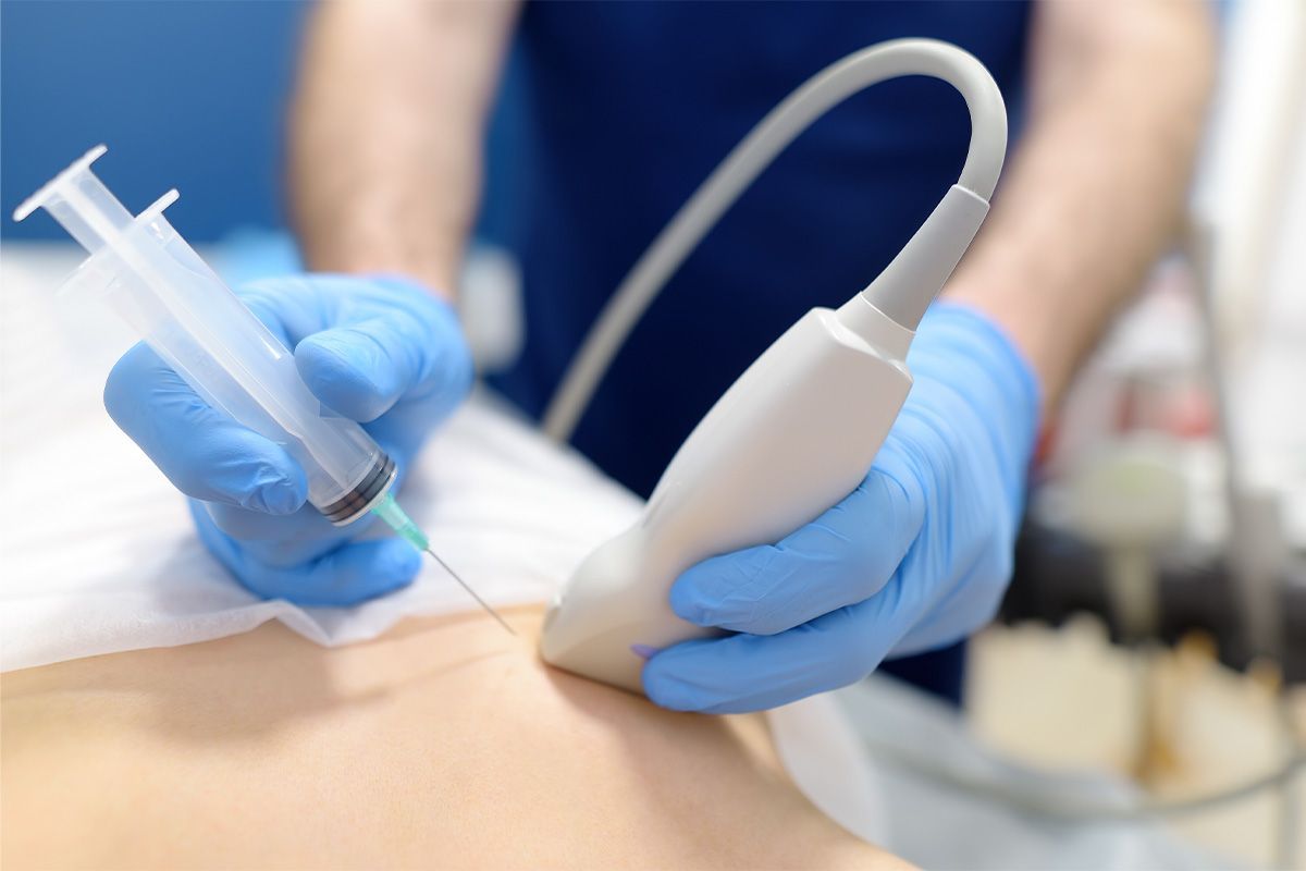
Image-guided biopsies use real-time imaging technology to guide a small needle to the area of concern. The goal is to remove a tiny tissue sample for further review.
How Imaging Technology Helps Guide the Biopsy Precisely
With traditional biopsies, a surgeon explores the area visually and by palpation, then takes a sample based on those findings. Image guidance, however, allows the radiologist to see exactly where the needle is in relation to the anomaly in the scan.
Why Image Guidance Reduces the Need for Surgical Biopsies
Imaging allows the radiologist to “see” through the skin without making a cut. That means:
- Less trauma to the body
- No scarring
- A faster, more accurate procedure
The Difference Between Image-Guided and Traditional Biopsies
| Feature | Traditional Surgical Biopsy | Image-Guided Biopsy |
|---|---|---|
| Anesthesia | General (Asleep) | Local (Numbed area) |
| Incision | Several centimeters; stitches | A few millimeters; bandage |
| Recovery | Days to weeks | 24–48 hours |
| Accuracy | High | Extremely high (Targeted) |
Types of Image-Guided Biopsies Performed at NVRA
The radiologists at NVRA have several imaging options available for biopsies. Depending on the location and nature of the anomaly, they will choose the best method for each patient's needs.
Ultrasound-Guided Biopsies and When They’re Used
This is the most common method for image-guided biopsies done at NVRA. It has the advantage of not exposing the patient to any level of radiation. Ultrasounds are a quick and comfortable option for patients and are frequently used for biopsies of:
- Breast lumps
- Thyroid nodules
- Lymph nodes
MRI-Guided Breast Biopsies for Hard-to-See Findings
Some anomalies are only visible with an MRI. For these patients, our board-certified physician uses specialized MRI equipment to target the area of interest. It is the most precise option for patients with high-risk factors or complex findings.
How Providers Determine the Best Imaging Method for Each Case
The choice depends on which view provides the clearest path to the tissue. At NVRA, our subspecialized radiologist will review prior scans to determine the most precise approach.
What an Image-Guided Biopsy Can Detect
The role of an image-guided biopsy is to collect a sample. The sample is sent to a pathologist, a specialist who examines the tissue under a microscope to provide a definitive diagnosis or rule it out.
How Biopsies Help Identify Benign Versus Malignant Conditions
The pathologist looks for patterns at the cellular level. That examination allows them to determine if the cells are:
- Atypical – not quite normal but not cancerous
- Malignant – cancerous
- Benign – non-cancerous and noninvasive
Inflammation, Infection, and Non-Cancerous Tissue Changes
People tend to associate a biopsy with cancer, but it can also detect:
- Infections – specific bacteria or fungi can cause a mass
- Fat necrosis – firm lumps that appear after an injury to fat tissue
- Autoimmune issues – some systemic conditions can cause localized inflammation
Why Biopsy Results Help Guide Next Steps in Care
A biopsy produces a pathology report that your care team can use as a roadmap. Benign results typically require nothing but annual screening. If the biopsy shows a pre-cancerous or cancerous condition, it will specify the type and grade, allowing your doctor to create a treatment plan.
What to Expect During Your Image-Guided Biopsy
Knowing what to expect from an image-guided biopsy will take some of the mystery out of it.
Step-by-Step Overview of the Procedure
- Positioning: A technician will help position you comfortably on an exam or specialized biopsy table.
- Numbing: The radiologist will clean the skin around the biopsy area and apply a local anesthetic to numb it.
- Targeting: Targeting is set up using the proper imaging tool, usually an ultrasound or MRI.
- Sampling: The radiologist will insert a small needle into the targeted location. You may feel pressure, but there should be no pain. The guided needle takes a small piece of tissue for examination.
- Marker Placement: Typically, a small, harmless metallic marker is left at the site of the biopsy so it can be identified on future scans if necessary.
- Completion: Pressure on the area helps to prevent bruising, and a small bandage is all that is needed to cover it. There are no stitches.
How Long the Biopsy Typically Takes
The procedure will take from 15 to 30 minutes. There may be additional time for preparation and post-care instruction. In most cases, you will be in and out within an hour.
Understanding Your Biopsy Results
The results typically take three to seven business days, or more, depending on the lab, to finalize. Your physician will receive a detailed report from the pathologist and go over the findings with you. They will explain how those findings relate to the original imaging.
Why Patients Choose NVRA for Image-Guided Biopsies
At Naugatuck Valley Radiological Associates, we understand the anxiety that comes with a biopsy. We combine the latest technology with our patient-centered approach to make the experience as stress-free as possible.
For 35+ years, our goal has been to provide the highest level of precision. We will answer any questions you may have about the procedure to help you feel more comfortable.
Image-guided biopsies are available at our three convenient NVRA locations. Check availability here or contact us to schedule your appointment.