When Is an Arthrogram Needed? Understanding MRI and CT Arthrography in Joint Pain Diagnosis
If you’ve been dealing with lingering joint pain, especially in your shoulder, hip, knee, or wrist, and traditional scans haven’t given clear answers, your provider may recommend an arthrogram. While the word might sound a little intimidating, the procedure itself is safe, targeted, and incredibly useful for diagnosing joint problems that are easy to miss on standard imaging.
At Naugatuck Valley Radiological Associates (NVRA), we perform both MRI with arthrogram and CT arthrogram exams, depending on what your doctor is looking for. This guide will walk you through what arthrography is, why it’s used, how the tests differ, and what you can expect if you’re scheduled for one.
What Is an Arthrogram?
Defining Arthrography in Simple Terms
An arthrogram is a specialized imaging test used to examine joints more closely, particularly when other tests, such as a standard MRI or X-ray, do not provide a clear explanation for ongoing pain or mobility issues. It combines contrast dye with either MRI or CT imaging to give a clearer view of the soft tissues inside a joint, like ligaments, cartilage, tendons, and the joint capsule.
Unlike basic scans, arthrography allows radiologists to spot small tears, structural damage, or inflammation that may be missed otherwise.
How It Differs from a Standard MRI or CT Scan
While a standard MRI or CT can show the general structure of bones and soft tissue, an arthrogram adds contrast directly into the joint, helping to highlight movement and structure in a more detailed way. This is especially helpful when assessing joint injuries, surgical complications, or unexplained mechanical symptoms, such as locking or catching.
When Do Doctors Recommend an Arthrogram?
Unexplained or Persistent Joint Pain
If you’ve had joint pain that hasn’t resolved with treatment — or previous scans were inconclusive — your provider may refer you for an arthrogram to get more answers. This is common in athletes, post-surgical patients, and individuals with long-term joint instability.
Evaluating Ligaments, Tendons, and Cartilage
Arthrography is particularly useful when your physician suspects injury or damage to internal joint structures that can’t be seen well on routine imaging. This includes labral tears in the shoulder or hip, meniscus injuries in the knee, or ligament damage in smaller joints, such as the wrist.
When Other Imaging Doesn’t Provide Enough Detail
Sometimes a standard MRI or CT scan is perfectly appropriate. However, in cases where pain persists without a clear cause or surgery is being considered, the added precision of an arthrogram can significantly impact diagnosis and treatment planning.
MRI Arthrogram vs. CT Arthrogram
How Each Test Works
At NVRA, MRI and CT arthrograms begin with a contrast injection directly into the joint. Instead of using fluoroscopy, we now perform this step using our CT scanner for image guidance. This allows our radiologists to precisely place the contrast agent using real-time CT imaging, ensuring accuracy and patient comfort.
Once the injection is complete:
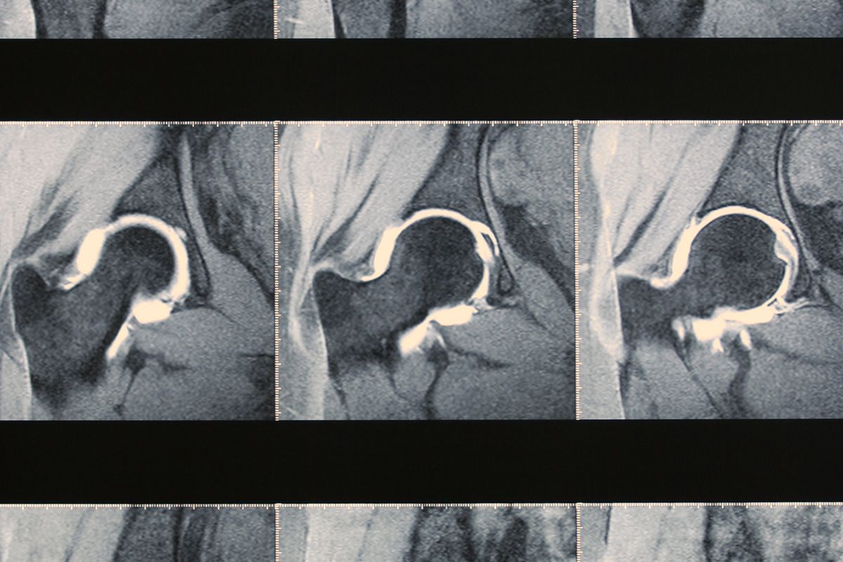
- An MRI arthrogram uses a magnetic field and radio waves to produce highly detailed images of soft tissues like cartilage, tendons, and ligaments.
- A CT arthrogram uses X-ray technology to create high-resolution images that show both soft tissues and bone structures with excellent clarity.
Key Differences Between MRI and CT Arthrography
| Feature | MRI Arthrogram | CT Arthrogram |
|---|---|---|
| Best For | Soft tissue detail (e.g., cartilage, labrum, tendons) | Bone detail and joint space evaluation |
| Radiation | None | Yes (low dose) |
| Scan Time | Longer (30–45 min) | Shorter (10–15 min) |
| Noise/Comfort | Louder, enclosed space | Quieter, shorter scan |
Your doctor will determine which test is right for your case depending on your symptoms, medical history, and the joint involved.
Which Joints Are Typically Imaged
NVRA commonly performs arthrograms on the following joints:
- Shoulder – Labral or rotator cuff tears
- Hip – Labral injuries or impingement
- Knee – Meniscus and ligament damage
- Wrist – TFCC tears or instability
- Elbow and ankle – Less frequent, but possible in certain cases
What Patients Can Expect During the Procedure
The Role of Contrast Dye in Arthrography
Contrast dye makes the internal structures of your joint more visible by creating separation between soft tissues. It’s injected into the joint before the imaging portion begins and usually contains a mix of saline and gadolinium (for MRI) or iodine-based contrast (for CT). Most patients tolerate it very well.
Step-by-Step Experience of the Test
- Prep: You’ll be positioned on the exam table. The skin is cleaned and numbed at the injection site.
- Injection: A radiologist uses real-time imaging to guide the contrast dye into the joint.
- Scan: You’ll move into the MRI or CT scanner immediately after the injection.
- Post-Scan: The entire process takes about 45–60 minutes. You may feel mild pressure, but most patients describe the procedure as tolerable.
Comfort, Safety, and Recovery Considerations
- You can resume normal activities the same day unless otherwise directed.
- Mild soreness is normal for a few hours after the procedure.
- Complications are rare but may include temporary swelling or mild discomfort at the injection site.
Benefits of Arthrography in Diagnosing Joint Pain
Earlier and More Accurate Diagnoses
The added clarity from an arthrogram can help identify conditions earlier — especially in complex or chronic joint issues — allowing your care team to move forward with confidence.
Better Treatment Planning and Surgical Decisions
Whether you’re considering physical therapy, injections, or surgery, arthrography helps guide your next steps by providing your orthopedic specialist with the most complete picture possible.
Helping Patients Understand Their Condition
A better image means a better conversation with your provider. Identifying the exact cause of your joint pain enables more precise explanations, stronger patient-physician communication, and more personalized care.
Potential Risks and Considerations
While arthrography is a very safe test, there are still a few things to be aware of:
- Mild discomfort or stiffness for 24–48 hours
- A small risk of infection or allergic reaction to contrast (extremely rare)
- Not suitable for patients with specific contrast allergies or kidney issues
Your doctor will always weigh the benefits and risks before recommending an arthrogram, and our team at NVRA ensures you’re fully informed before your scan.
How Arthrography Fits Into Your Overall Care Plan
An arthrogram isn’t always the first imaging test — but when joint pain doesn’t respond to basic imaging or treatment, it can be the most helpful next step. It’s beneficial for planning orthopedic surgery or ruling out minor but important injuries that might otherwise go undetected.
Advanced Joint Imaging at NVRA
At Naugatuck Valley Radiological Associates, we’re committed to supporting patients and referring providers with advanced imaging, prompt results, and a personalized experience. If you’ve been referred for an MRI or CT arthrogram, our team is ready to walk you through each step with clarity, comfort, and compassion.
Arthrography is available at select NVRA locations — check availability here or contact us to schedule your appointment.