Diagnostic Imaging for Back Pain: MRI, CT, or X-Ray?

“Oh, my aching back!”
If you are like most people, you will be uttering those words at some time in your life. In fact, a national survey shows that 39% of people in the United States had back pain within the previous three months; up to 80% of Americans will experience back pain at some point during their lives. In fact, low back pain is the second most common health complaint in the United States; 80 to 85% of people experience low back pain at some time.
Having back pain is no fun, and neither is wondering why your back is hurting. Diagnostic imaging may be helpful, but determining when to undergo imaging for back pain can be challenging. When you and your doctor decide that it is time for diagnostic imaging, NVRA is glad to provide the imaging you need for your back pain.
Diagnostic Imaging for Back Pain: When to Consider an MRI, X-Ray, or CT Scan
Several conditions can cause back pain. Treatment for these conditions relies largely on the underlying cause.
Back Pain and Diagnostic Imaging
Diagnostic imaging can help you and your doctor determine what is causing your back pain.
Common Causes of Back Pain
- Strains – injuries to your muscles or tendons, which are the tissues that attach muscles to bones; strains can happen suddenly or over time.
- Sprains – injuries to the ligaments that connect the bones of the spine, known as vertebrae; spinal sprains usually occur after a fall, twisting motion, or blunt force injury.
- Fractures – broken bones of the spine.
- Arthritis – pain, swelling, stiffness of the joints between vertebrae.
- Degenerative disk disease – thinning of the rubbery discs that cushion the vertebrae and prevent the bones from grinding together.
- Slipped disk – also known as a herniated disk, a slipped disk occurs when the disk tears, and the gel-like center of the disk pushes through the crack to press against nearby nerves to cause pain.
- Osteoporosis – loss of bone density or weakening of the bones.
- Sacroiliitis – inflammation and irritation of the sacroiliac joint, which is where the spine connects to the pelvis.
- Spinal stenosis – abnormal narrowing of the space inside the spinal column, which puts pressure on the spinal cord to cause pain, numbness, tingling, or weakness.
- Curvature of the spine – abnormal curves of the spine, such as scoliosis (sideways curvature), kyphosis (curvature of the upper back that causes a hump), or lordosis (excessive inward curvature of the lower back).
Less common causes of back pain include:
- Tumors
- Spina bifida – a birth defect in which the vertebrae and spinal cord do not form correctly
- Multiple sclerosis – a condition in which the immune system attacks the covering that protects nerves
Importance of Accurate Diagnosis
Because so many things can cause back pain, and because each underlying cause of back pain requires a different approach to treatment, diagnosing back pain can be complicated. Strains and sprains usually respond well to rest and over-the-counter pain relievers, for example, while a spinal fracture may require surgery. Knowing the cause of your back pain can help you and your doctor determine the best course of treatment.
Overview of Imaging Modalities for Back Pain
Doctors rely on different types of imaging tests, known as imaging modalities, for diagnosing back pain. Diagnostic tests for lower back pain include X-rays, CT scans, and MRIs. Each imaging modality provides different insight into back pain.
X-Ray Imaging
Because they are fast and inexpensive, X-rays are usually the first type of imaging done for back pain. Doctors typically recommend X-ray imaging for backs when a patient:
- Has recently had an injury
- Is experiencing pain that does not respond to conservative treatment, such as rest, ice, and physical therapy
- Has pain along with other symptoms
- Has “red flags,” such as a fever, weight loss, history of cancer, or neurological symptoms, such as tingling, numbness, or weakness
X-ray imaging can show:
- Fractures
- Osteoporosis
- Arthritis
- Tumors
- Slipped discs
Computed Tomography (CT) Scans
CT scans create 3D images of your spine. It uses an X-ray and a computer to compile multiple images into 3D images. Your doctor may recommend a CT for back pain when an X-ray or a physical examination is inconclusive.
CT scans of the spine can show:
- Herniated disks
- Injuries
- Spina bifida
- Fractures
- Arthritis
- Tumors
- Spinal stenosis
Magnetic Resonance Imaging (MRI)
MRIs use a strong magnetic field and radio waves to create images of the spine. When it comes to back pain, this imaging modality has several advantages over X-rays and CT scans. For example, an MRI is the most effective imaging method for creating images of the spinal cord and nerves.
When Is Diagnostic Imaging Needed?
Because back pain usually goes away on its own within a few weeks, doctors usually suggest waiting 6 weeks before undergoing diagnostic imaging for most patients. In many cases, diagnosing back pain can be done through a physical examination.
Scenarios Requiring Immediate Imaging
Some patients require immediate imaging. A doctor might recommend imaging right away if a patient has:
- Severe neurological problems, such as weakness, numbness, or bowel/bladder issues, or neurological problems that get worse
- Fever
- Sudden back pain with tenderness of the spine, especially in patients with a history of weak bones (osteoporosis), cancer, or steroid use
- Trauma
- A serious underlying medical condition, such as cancer
Why MRI for Spine Assessment?
Your doctor may order an MRI first, or they may recommend having an MRI for spine pain after other modalities, such as an X-ray or CT scan.
What does an MRI show for back pain?
An MRI can show a number of conditions, such as herniated disks, spinal stenosis, nerve compression, and other painful conditions.
Advantages of MRI Over Other Modalities
MRI has certain advantages over X-rays and CT scans, making it the preferred scan for back pain in many cases. For example, CT scans and X-rays use small doses of radiation to create images, whereas MRIs do not.
MRI vs. CT for back pain
MRI is generally better for creating images of soft tissue, such as the spinal cord, nerves, and herniated disks. CT is good for visualizing the structure of bones and for detecting bone fractures.
Detailed Visualization of Soft Tissues and Nerves
X-rays and CT scans are very effective at creating images of bones and empty spaces, and they can do a fair job of visualizing some types of soft tissue. However, MRIs create highly detailed images of soft tissues and nerves, which may be important in diagnosing the underlying cause of back pain.
Preparing for a Spinal MRI
Preparing for a spinal MRI may be easier than you think:
- Eat and drink as usual, unless directed otherwise
- Take all medications as usual
- Remove all piercings and leave all jewelry at home
What to Expect During the MRI
Prior to your MRI, the radiology team will likely have you change into a hospital gown and confirm that you have no metal on the inside or outside of your body.
Reading and Understanding MRI Results
A radiologist will interpret the MRI results and issue a report, which usually includes different sections for patient information, details about how the MRI was performed, the findings, and the radiologist’s impressions/conclusion. The findings section includes information about the structures examined, along with abnormalities, such as disk herniations or spinal stenosis. The impression/conclusion provides a summary of the findings and their significance.
Interpreting Normal Versus Abnormal Findings
Normal findings show a clear spinal canal, healthy vertebrae, and intact disks. Abnormal findings may show impingement or entrapment (pinched nerve), degenerative disk disease, bulging of the disks, fractures, improper development of the spinal column, inflamed tissue near the spine, and more.
Common Conditions Diagnosed Through MRI
MRIs can help doctors diagnose a number of conditions that cause back pain, such as:
- Tumors
- Abscesses (infections)
- Birth defects
- Bleeding into the spinal cord
- Degenerative disk disease
- Multiple sclerosis
- Inflammation of the spinal cord
MRI can also help surgeons plan surgical procedures.
Potential Limitations and Risks of MRI
While MRIs can create incredibly clear images of the spine, muscles, and other structures of the back, the imaging modality does have some potential limits and risks.
Recognizing the Limitations of MRI Scans
MRIs are also limited in their ability to image bones and other tissues in your back.
Potential Risks and Precautions
MRI scans use a strong magnetic field to create images, and this magnetism can affect implanted devices (such as pacemakers) or move metallic objects. Be sure to tell the radiation technician if you have any implanted metal or are wearing metal jewelry.
Patient Education and Expectations
Patients often don’t know what to expect with an MRI. They are usually relieved to learn that having an MRI is relatively easy.
Do I need an MRI for back pain?
You may need an MRI for back pain that has persisted longer than 6 weeks, if you have recently suffered trauma to your back, or if you have a “red flag,” such as numbness or cancer.
Educating Patients About MRI Processes
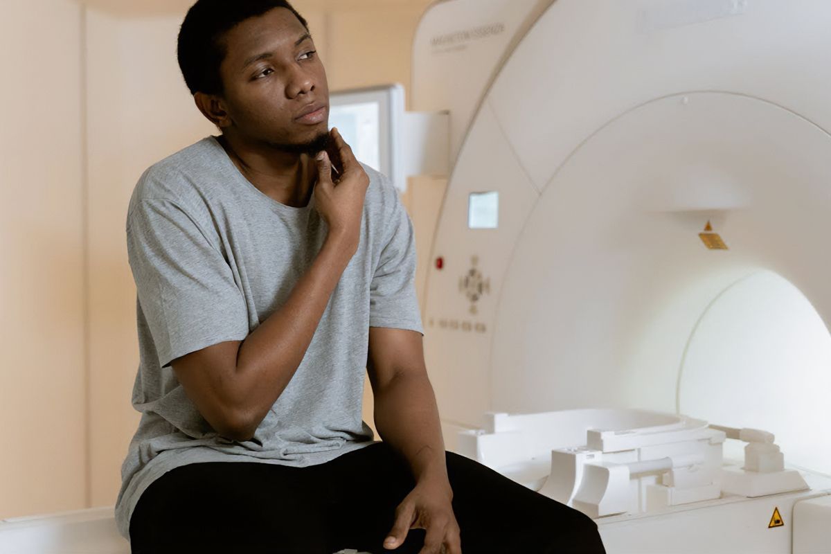
Prior to your MRI, the radiology team will instruct you to lie on your back on a table. This table will slide into the MRI machine, which looks like a long tube that is open at both ends. During the test, the MRI is quite loud, so the team may provide you with a set of headphones and music that helps drown out the noise. Doctors will sometimes order contrast that highlights certain features during the test; in these cases, the radiology team will start an IV.
You will need to lie perfectly still during your MRI to prevent the images from being blurry. If you have trouble lying still, or if you experience anxiety or other discomfort, you can alert the technologist by pressing an alarm button.
Your MRI results should be available within a few days. To learn the results of your MRI, please contact the physician who ordered them.
For more information about when to consider an MRI, X-ray, or CT scan for back pain, contact Naugatuck Valley Radiology. We offer a wide variety of modalities and imaging for back pain.


